Back Human Bones Labeled - Human Skeleton In Front Profile And Back Views With Main Parts Labeled Vector Illustration In Flat Style Over White Background Stock Vektorgrafik Adobe Stock - Each lumbar spinal level is numbered from top to bottom—l1 through l5, or l6.
Back Human Bones Labeled - Human Skeleton In Front Profile And Back Views With Main Parts Labeled Vector Illustration In Flat Style Over White Background Stock Vektorgrafik Adobe Stock - Each lumbar spinal level is numbered from top to bottom—l1 through l5, or l6.. Below the lumbar spine is a bone called the sacrum, which makes up the back part of the pelvis. Human backbone diagram, bone, human backbone diagram. Back human bones labeled : The upper cervical region (c1 and c2), and the lower cervical region (c3 through c7). The range of motion in the thoracic spine is limited.
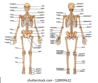
The axial skeleton and the appendicular skeleton. Although the cranium—the largest section of the skull—might appear to be one solid bone, there are actually 22 bones that encase the brain. Vertebrae, bones, joints, ligaments, muscles, muscular system, fascia, arteries, veins, nerves and various adjacent organs. Human body muscles human body organs human body parts human organ diagram body organs diagram anatomy organs anatomy bones heart anatomy body muscle anatomy. The lumbar spine is the lower back that begins below the last thoracic vertebra (t12) and ends at the top of the sacral spine, or sacrum (s1).
Human Skeleton High Res Stock Images Shutterstock from image.shutterstock.com Upper back anatomy bones : When autocomplete results are available use up and down arrows to review and enter to select. The upper cervical region (c1 and c2), and the lower cervical region (c3 through c7). The twelve thoracic vertebrae are numbered t1 to t12. See human skull anatomy stock video clips. To learn all about the skeleton system in the human body, check out this guide. The vertebral column of the lower back includes the five lumbar vertebrae, the sacrum, and the coccyx. Human anatomy arm bones from atlas 12 photos of the human anatomy arm bones from atlas , bone.
The axial skeleton and the appendicular skeleton.
The axial skeleton and the appendicular skeleton. Bone (os frontale), which already belongs to the neurocranium, at the top: Throughout the spine, intervertebral discs made of. When autocomplete results are available use up and down arrows to review and enter to select. Facet joints connect each vertebra, with fluid supporting. Vertebrae, bones, joints, ligaments, muscles, muscular system, fascia, arteries, veins, nerves and various adjacent organs. Upper back anatomy bones : The lumbar spine is the lower back that begins below the last thoracic vertebra (t12) and ends at the top of the sacral spine, or sacrum (s1). These bones work together to provide flexibility to the trunk, support the muscles of the trunk, and protect the spinal cord and spinal nerves of the back. To learn all about the skeleton system in the human body, check out this guide. See human skull anatomy stock video clips. Your skeleton can be divided into two main parts. Human body muscles human body organs human body parts human organ diagram body organs diagram anatomy organs anatomy bones heart anatomy body muscle anatomy.
To learn all about the skeleton system in the human body, check out this guide. The part of the nerve that emerges out of the spine is called the nerve root. Superficial back muscles, intermediate back muscles and intrinsic back muscles.the intrinsic muscles are named as such because their embryological development begins in the back, oppose to the superficial and intermediate back muscles which develop elsewhere and are therefore classed as extrinsic muscles. Human body anatomy female female anatomy muscle shoulder blade pain anatomy back muscles bones man female anatomy body muscles in a body female anatomy muscole shoulder concept muscular sysyem. Upper back anatomy bones :
What Is A Skeletal System Diagram With Pictures from images.infobloom.com Human body muscles human body organs human body parts human organ diagram body organs diagram anatomy organs anatomy bones heart anatomy body muscle anatomy. The cervical spine is further divided into two parts; The lumbar spine is the lower back that begins below the last thoracic vertebra (t12) and ends at the top of the sacral spine, or sacrum (s1). Each lumbar spinal level is numbered from top to bottom—l1 through l5, or l6. The range of motion in the thoracic spine is limited. Throughout the spine, intervertebral discs made of. The central feature of the human back is the vertebral column, specifically the length from the top of the thoracic vertebrae to the bottom of the lumbar vertebrae, which houses the spinal cord in its spinal canal, and which generally has some curvature that gives shape to the back. It is composed of 300 bones at birth, but later decreases to 80 bones in the axial skeleton and 126 bones in the appendicular skeleton.
Medical vector illustration isolated on white background.
See human back anatomy stock video clips. Spine or vertebral column | spine bones joints | human spine anatomy 3d animation | elearninthis video illustrates one of the main parts of human body, the s. The axial skeleton and the appendicular skeleton. The lumbar spine is the lower back that begins below the last thoracic vertebra (t12) and ends at the top of the sacral spine, or sacrum (s1). The axial skeleton is made up of the skull, backbone, breastbone, and ribs. When humans are born we have close to 300 bones, and over time they fuse together. It is composed of 300 bones at birth, but later decreases to 80 bones in the axial skeleton and 126 bones in the appendicular skeleton. The twelve thoracic vertebrae are numbered t1 to t12. Nerves in your lower back five pairs of lumbar spinal nerves labeled l1 to l5 branch off your spinal cord and exit through small holes between the vertebrae. See human skull anatomy stock video clips. See sacrum (sacral region) the sacrum is connected to part of the pelvis (the iliac bones) by the sacroiliac joints. Skeleton human skeleton labelled skeleton skeleton labelling skeleton label bones skeleton powerpoint the human skeleton the skeleton bones are crucial to enabling movement about joints, too. Throughout the spine, intervertebral discs made of.
Facet joints connect each vertebra, with fluid supporting. Superficial back muscles, intermediate back muscles and intrinsic back muscles.the intrinsic muscles are named as such because their embryological development begins in the back, oppose to the superficial and intermediate back muscles which develop elsewhere and are therefore classed as extrinsic muscles. Touch device users, explore by touch or with. This bone is shaped like a triangle that fits between the two halves of the pelvis, connecting the spine to the lower half of the body. On anatomical parts the user can choose to display the various structures in colored illustrations of the anatomy of the back and spine:
Flat Bones Definition Examples Diagram And Structure from post.healthline.com Human backbone diagram, bone, human backbone diagram. The range of motion in the thoracic spine is limited. It is composed of 300 bones at birth, but later decreases to 80 bones in the axial skeleton and 126 bones in the appendicular skeleton. Bone structure of leg, above and below 12 photos of the bone structure of leg, above and below , bone. 12 photos of the human back bone chart. Upper back anatomy bones : Medical vector illustration isolated on white background. Back human bones labeled :
These bones work together to provide flexibility to the trunk, support the muscles of the trunk, and protect the spinal cord and spinal nerves of the back.
When humans are born we have close to 300 bones, and over time they fuse together. Medical vector illustration isolated on white background. See sacrum (sacral region) the sacrum is connected to part of the pelvis (the iliac bones) by the sacroiliac joints. C1 is termed the atlas and c2 the axis. Human back bone chart, find out more about human back bone chart. Quizzes on human skeletal system anatomy, bone anatomy, and bone markings. It is composed of 300 bones at birth, but later decreases to 80 bones in the axial skeleton and 126 bones in the appendicular skeleton. Facet joints connect each vertebra, with fluid supporting. The humerus is divided into 3 parts, the round head, the narrow neck, and the tubercles. Skeleton human skeleton labelled skeleton skeleton labelling skeleton label bones skeleton powerpoint the human skeleton the skeleton bones are crucial to enabling movement about joints, too. The part of the nerve that emerges out of the spine is called the nerve root. On anatomical parts the user can choose to display the various structures in colored illustrations of the anatomy of the back and spine: The upper cervical region (c1 and c2), and the lower cervical region (c3 through c7).
The twelve thoracic vertebrae are numbered t1 to t12 human back bones. Facet joints connect each vertebra, with fluid supporting.


0 Komentar